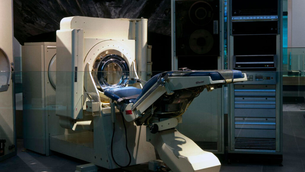
In the annals of medical history, a breakthrough occurred 50 years ago that forever altered the landscape of diagnostic imaging. The date was September 1, 1973, and the world bore witness to a remarkable advancement in medical science: X-rays, traditionally used for capturing static images of bones, delved deep into the mysteries of the living brain. This groundbreaking moment was captured in a headline: “X-rays of the brain in thin cross sections — Science News.”
The catalyst for this transformative leap was an innovative device, celebrated by physicians as a watershed moment in the realm of X-ray diagnosis. It promised to unveil the innermost recesses of the brain, offering an unprecedented level of detail while subjecting patients to minimal radiation exposure. This device, known as the EMI scanner, marked the birth of X-ray computed tomography (CT) and set in motion a revolution in medical imaging.
The Birth of X-ray Computed Tomography
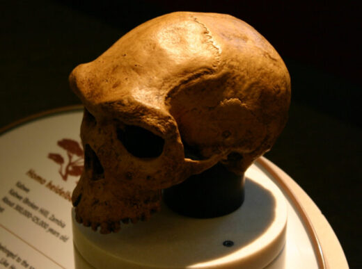
Fast forward to today, and CT scanning stands as a cornerstone of modern medicine, enabling physicians and researchers to peer not only into the intricate complexities of the human brain but also to explore the inner workings of various organs, scrutinize bone structures, and even navigate the intricate network of blood vessels. The evolution of CT technology has not only transformed the field of medicine but has also found applications in diverse scientific domains, from archaeology to zoology.
A Crossroads of Discovery: CT Scanning Beyond Medicine
While the roots of CT scanning lie in medical diagnosis, its branches have extended far beyond the realm of healthcare. It has become an invaluable tool for scientific exploration across numerous disciplines. Consider, for instance, the world of zoology, where CT scanning has helped unlock the secrets of creatures great and small. In the case of pumpkin toadlets, these diminutive amphibians posed an enigmatic question: Why were they such clumsy hoppers? Through the lens of CT scanning, researchers ventured into the inner ears of these toadlets, revealing a potential answer—tiny inner ears that might hinder their ability to maintain balance.
The Dawn of Precision: Photon-Counting CT Scanners
Innovation continues to propel CT scanning into new frontiers. A recent milestone came in the form of “photon-counting” CT scanners, earning approval from the U.S. Food and Drug Administration (FDA) last year. These cutting-edge scanners promise to elevate imaging precision to unprecedented heights. With sharper images and enhanced capabilities, photon-counting CT scanners are poised to unravel previously inscrutable scientific enigmas.
Closing Thoughts: A Journey from the Past to the Future
As we commemorate the 50th anniversary of that momentous day in 1973 when X-rays pierced the shroud of mystery surrounding the human brain, we celebrate not only the strides made in medical imaging but also the indomitable spirit of exploration that continues to drive scientific progress. CT scanning has transcended its original purpose, becoming a cornerstone of medical diagnosis and a guiding light for diverse scientific disciplines.
As we gaze back at the journey from the birth of X-ray computed tomography to the emergence of photon-counting CT scanners, we stand at the precipice of ever greater discoveries. These innovations exemplify the boundless potential of human ingenuity and the unyielding pursuit of knowledge.


